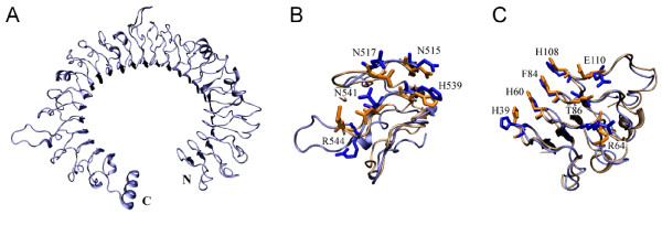
Figure 3.
Comparison of model and crystal structure of mouse TLR3 ectodomain at the two ligand interaction regions. Blue: structure obtained by homology modeling; orange: crystal structure (PDB code: 3CIG). (A) The modeled backbone structure of mouse TLR3 ectodomain. (B) Model and crystal structure superimposed at the N-terminal interaction region. The root mean square deviation is 1.96 Å. (C) Superimposition at the C-terminal interaction region. The root mean square deviation is 1.9 Å. The reported interacting residues are presented with side chain and labelled with residue name and position in (B) and (C).

