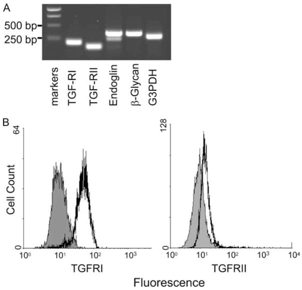
Figure 1.
Expression of TGF-β receptor in human skin mast cells. A, RT-PCR. mRNAs for TGF-RI (213 bp), TGF-RII (158 bp), endoglin (378 bp), β-glycan (364 bp), and G3PDH (303 bp) were detected by RT-PCR in skin-derived mast cells. B, Expression of TGF-RI and TGF-RII. Skin mast cells were incubated with mouse anti-TGF-RII mAb (black) or isotype control IgG (gray), followed by FITC-conjugated rat anti-mouse IgG. For TGFRI, cells were first fixed and permeabilized, and then incubated with either rabbit IgG (gray) or rabbit anti-TGFRI Ab (black), which recognizes the intracellular portion of TGF-RI. Alexa Fluor 488-conjugated goat anti-rabbit IgG was used as the secondary Ab. One representative experiment of three with different samples is shown in A and B.

