Figure 2.
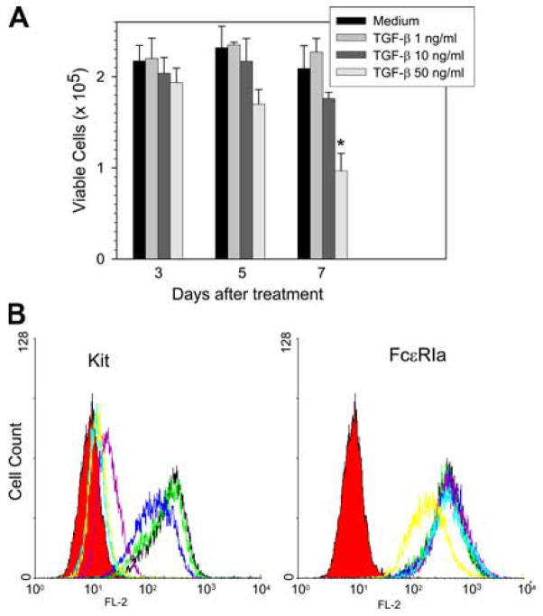
Effect of TGF-β on the viability of and surface expression of Kit and FcεRI on skin mast cells. A, Cell viability. Human skin mast cells were incubated with the indicated concentrations of TGF-β for up to 7 days. Viable cells were counted on days 3, 5, and 7 using trypan blue exclusion. Data are presented as mean ± SE of three independent experiments, each performed in duplicate. *, p < 0.05 compared with control group. B, Flow cytometry. Mast cells were incubated with TGF-β at 0 (black), 0.01 (green), 0.1 (blue), 1 (purple), 10 (light blue), and 50 (yellow) ng/ml for 3 days along with fresh medium containing SCF (100 ng/ml). Cells were then harvested and incubated with PE-conjugated mouse anti-CD117 or isotype IgG mAbs. FcεRI was labeled with 22E7 and PE rat anti-mouse IgG. Isotype controls are in red. Data are representative of three separate experiments with similar results.

