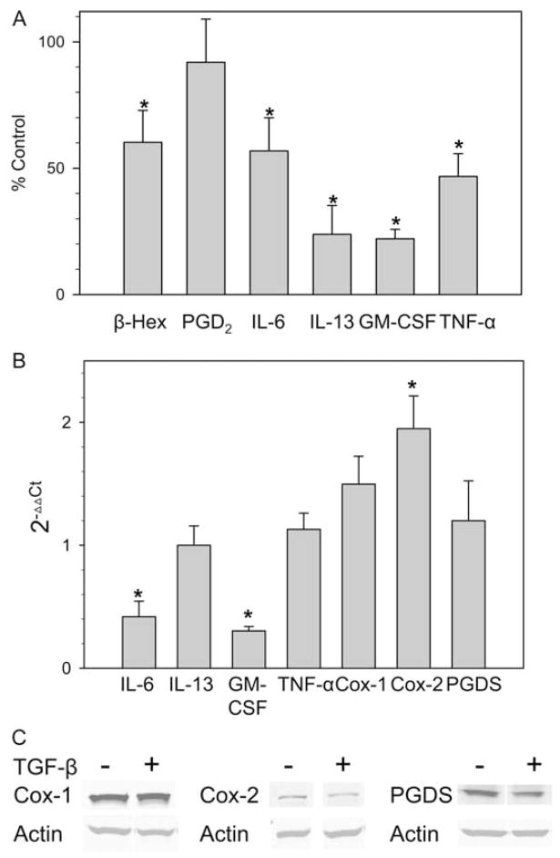
Figure 3.
TGF-β selectively regulates mediator release by FcεRI-stimulated skin mast cells. A, Production of cytokines and PGD2 by activated human skin mast cells. Mast cells were treated with TGF-β at 10 ng/ml for 2 days in the presence of 100 ng/ml SCF. The cells were washed in medium and activated with 1 μg/ml 22E7 mAb at 37°C. β-Hexosaminidase release and PGD2 and cytokine production were measured as described after 30 min, 45 min, and 24 h of stimulation, respectively. Data are displayed as mean ± SE values for cells treated with TGF-β as a percentage of control cells not exposed to TGF-β (n = 4 independent experiments for β-hexosaminidase and PGD2, n = 3 independent experiments for cytokine production). *, p < 0.05 compared with the control that was not exposed to TGF-β. B, Cellular mRNA levels of Cox-1, Cox-2, PGD synthase, and cytokines. Skin mast cells were treated with TGF-β for 2 days and then stimulated with 1 μg/ml 22E7. Enzyme mRNA levels were measured before stimulation, and cytokine levels were measured 3 h after stimulation by real-time RT-PCR. The 2−ΔΔCt values for TGF-β-treated cells were then calculated. Data are expressed as mean ± SE of three independent experiments, each performed in triplicate. *, p < 0.05 compared with the control that was not exposed to TGF-β. C, Cellular protein levels of Cox-1, Cox-2, and PGD synthase. These enzyme levels were measured by Western blotting 2 days after treatment of the cells with TGF-β and compared with medium. The blot shown is representative of three independent experiments.

