Figure 9.
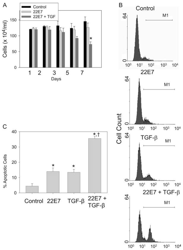
TGF-β enhances apoptosis of activated skin mast cells. Human skin mast cells were given fresh medium containing SCF alone, or simultaneously stimulated with 22E7 (1 μg/ml) ± TGF-β (10 ng/ml). A, Viable cell numbers. Cells were assessed by trypan blue exclusion on days 1, 3, 5, and 7 (mean ± SE, n = 3). *, p < 0.05 compared with medium control cells and to those activated with 22E7, but not treated with TGF-β. B, Detection of apoptotic mast cells. Skin mast cells at day 7 after being treated as in A were labeled with FAM-VAD-FMK and assessed by flow cytometry. Graphs represent results from one of three independent experiments. C, Percentage of apoptotic mast cells. Percentages of apoptotic cells at day 7 of three independent experiments, each performed in duplicate, as described in A and B (mean ± SE, n = 3). *, p < 0.05 compared with control group. †, p < 0.05 compared with 22E7-alone group or TGF-β-alone group.

