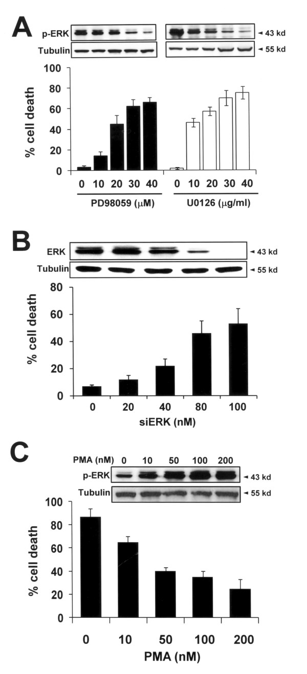
Figure 5.
Activation of ERK in HRT98G cells (A) HRT98G cells were pre-treated with the ERK inhibitor PD98059 or U0126 at the indicated concentration and a trypan blue exclusion assay was performed after 6 h of hypoxia. Immunoblots for p-ERK and tubulin are shown in the upper panel. (B) HRT98G cells were transfected with the indicated concentration of siERK for 48 h and cell death assay was performed as described in A. Immunoblots for ERK and tubulin are shown in the upper panel. (C) T98G cells were treated with the ERK activator PMA at the indicated concentration and cell death assay was performed as described in A. The immunoblots for p-ERK and tubulin are shown in the upper panel.

