
Fig. 1.
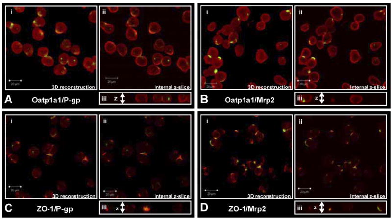
P-gp or Mrp2 staining with Oatp1a1 or ZO-1 in freshly isolated rat hepatocytes. i, three-dimensional reconstruction of z-stack (1-μm xy slices); ii, single internal 1-μM slice; and iii, cut-through of z-stack. A, Oap1a1 (red) and P-gp (green); B, Oatp1a1 (red) and Mrp2 (green); C, ZO-1 (red) and P-gp (green); D, ZO-1 (red) and Mrp2 (green).