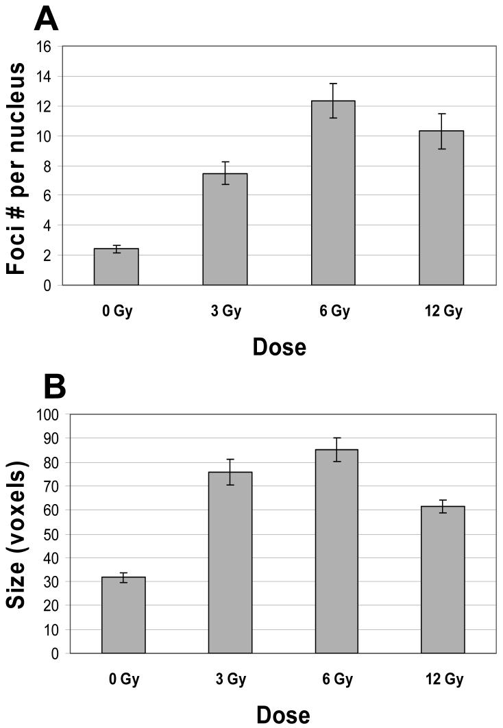
Figure 5.
(A): Induction of nuclear focus formation in human U-1 melanoma cells that were irradiated with 0 (unirradiated control), 3, 6, and 12 Gy of X-rays, then incubated at 37 °C for 4 h, fixed/permeabilized, and immunostained for Mre11 and Rad50 proteins, as described in Materials and Methods. As expected, 6 Gy and 12 Gy exposures induced more foci per nucleus than a 3 Gy exposure did. However, a 12 Gy exposure did not induce more foci per nucleus than a 6 Gy exposure. The differences between F values for 3 Gy-irradiated cells and F values for control (0 Gy) and 6 Gy-irradiated cells were statistically significant (P < 0.05). The differences between F values for 12 Gy-irradiated cells and F values for 3 Gy- and 6 Gy-irradiated cells were statistically insignificant (P > 0.05). The total numbers of analyzed nuclei of control cells (0 Gy), and 3 Gy-, 6 Gy-, and 12 Gy-irradiated cells, were 156, 79, 77, and 93, respectively. (B): The average size of nuclear foci in control cells (0 Gy), and in 3 Gy-, 6 Gy-, and 12 Gy-irradiated cells (shown in voxels). All doses caused an increase in the size of foci. The average size of nuclear foci in irradiated cells was in 1.9-2.7 times larger than the average size of those in control cells. The difference between the average size of nuclear foci in 12 Gy-irradiated cells and the average sizes of foci in 3 Gy- and 6 Gy-irradiated cells was statistically significant ( P < 0.05). Data presented are the mean ± standard error of the mean of five independent experiments.

