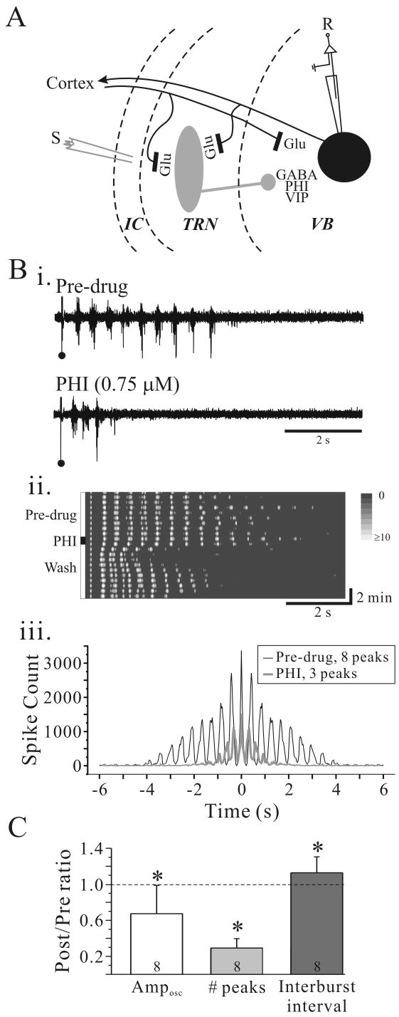
Figure 1.
PHI attenuates intrathalamic rhythmic activity. A. Simplified schematic illustrating thalamic circuitry with putative localization of PHI. Abbreviations: S, stimulus electrode; R, recording electrode; GABA, γ-aminobutyric acid; Glu, glutamate; PHI, peptide histidine isoleucine; VB, ventrobasal nucleus; TRN, thalamic reticular nucleus; IC, internal capsule. Bi. Extracellular multiple-unit recording from VB in a rat thalamic slice. In BMI (10 μM; Pre-drug), a single stimulus (•) in TRN evokes rhythmic discharge in VB. PHI (0.75 μM, 60 seconds) dramatically suppresses the rhythmic activity. Bii. Contour plot of experiment in Bi illustrates the time course of PHI effect on intrathalamic rhythmic activity. Prior to PHI application the rhythmic activity is very stable and lasts for many cycles. After PHI application, the rhythmic activity is dramatically attenuated, but returns near control levels within 5 minutes. Biii. Autocorrelogram of experiment in Bi illustrates a highly synchronized response that lasts nearly four seconds in control conditions (black trace). PHI (gray trace) reduces the numbers of peaks from 8 to 3. C. Summary of effects of PHI on oscillation amplitude (Amposc), number of peaks, and oscillation frequency. * p<0.05, **p<0.01.

