Figure 4.
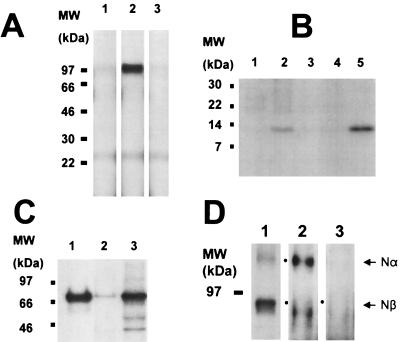
Processing of APP by M2 in HeLa cells. (A) SDS/PAGE patterns of immunoprecipitated APP Nβ-fragment (97-kDa band) from the condition media (2 h) of pulse–chase experiments. Lanes: 1, transfection of APP alone; 2, co-transfection of APP and M2; 3, same as lane 2 except that bafilomycin A1 is included. (B) SDS/PAGE patterns of APP βC-fragment (12 kDa) immunoprecipitated from the conditioned media of the same experiment as in A. Lanes: 1, transfection of APP; 2, co-transfection of APP and M2; 3, as in lane 2 but with bafilomycin A1; 4, transfection of Swedish APP; 5, co-transfection of Swedish APP and M2. (C) SDS/PAGE patterns of immunoprecipitated M2 (70 kDa). Lanes: 1, M2 transfected cells; 2, untransfected HeLa cells after long time film exposure; 3, endogenous M2 from HEK 293 cells. (D) SDS/PAGE patterns of APP fragments (100-kDa Nα-fragment and 95-kDa Nβ-fragment) recovered from conditioned media after immunoprecipitation using antibodies specific for the N-terminal region of APP. Lanes: 1, autoradiogram of immunoprecipitation from co-transfection of APP and M2; 2 and 3, immunoblotted by using antibody Aβ1–17, specific for the Nα-fragment that does not recognize the Nβ-fragment; 2, transfection with APP alone; 3, co-transfection of APP and M2. Dots mark band positions.

