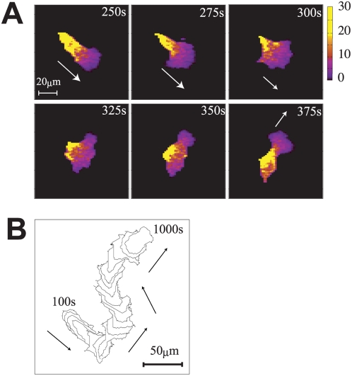
Figure 2. Simulated amoeboid-like locomotion.
(a) Snapshots of the distribution of the cortical factor in the
amoeboid-like locomotion. Parameters are set to  . Arrows in the panel indicate direction of motion of
the cell. Colors indicate the concentration of the cortical factor. At
the rear of the moving cell, the concentration of the cortical factor
often exceeds its equilibrium value
  . (b) A track of the amoeboid-like locomotion from 100
s to 1000 s drawn at every 50 s. The track at later steps masks the
track of earlier steps. Arrows indicate the direction of motion.

