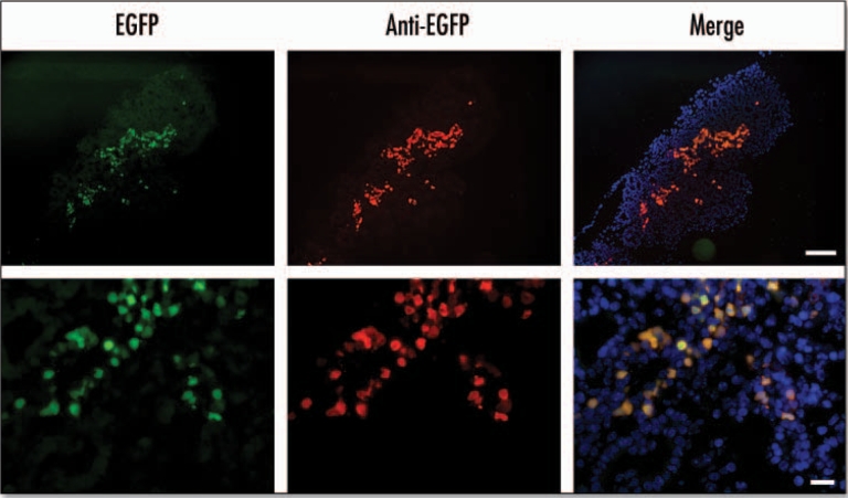
Figure 3.
EGFP fluorescence colocalizes with anti-EGFP staining in the E15.5 pancreas. In the panels marked EGFP the fluorescence was detected using EGFP filters. In panels marked anti-EGFP immunohistochemistry was performed on cryosections using an anti-EGFP antibody and a Texas Red conjugated secondary antibody with DAPI staining to identify nuclei. The merged image was obtained by merging images using Photoshop 6.0 software (Scale bar for: upper panels = 100 µm, lower panels = 20 µm).

