Figure 5.
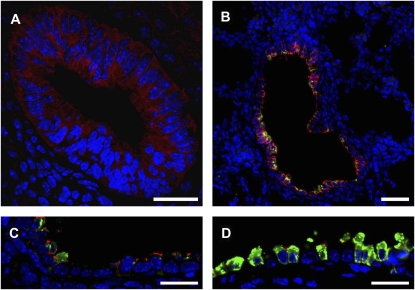
Claudin 10 is expressed early in mouse lung development. Mouse lungs from E14.55 or E18.5 embryos, or from P5 mice, were immunostained for localization of CCSP, Cldn10, and CGRP and images acquired by fluorescence microscopy. Nuclei were visualized using a DAPI nuclear counterstain (blue). (A) Cldn10 (red) is expressed throughout the developing airway tubules at E14.5. Scale bar = 25 μm. (B) Intense Cldn10 staining (red) is seen along lateral membranes of airway epithelial cells. Faint CCSP immunostaining (green) is seen within the airway at this developmental stage. Scale bar = 120 μm. (C) Tissue from E18.5 dpc embryos was stained as above. A row of four CCSP-negative, Cldn10-negative cells is seen surrounded by epithelial cell with strong Cldn10 lateral membrane staining and weak CCSP staining. Scale bar = 25 μm. (D) Lung tissue from a 5-day-old mouse was immunostained as above. CCSP expression has greatly increased, and Cldn10 staining remains restricted to CCSP immunoreactive cells. Scale bar = 25 μm.

