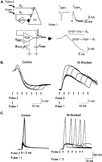
Figure 2. Wide dynamic range aVLSI microchip emulation of coincidence detection via feedforward inhibition.
(A) Network arrangement and synaptic compartment. Left top: A hippocampal network architecture (Pouille, 2001) of Schaffer collateral inputs (S1 and S2) on to a pyramidal cell (PC); IN–interneuron; ●–inhibitory synapse; ◁–excitatory synapse. Left bottom: A membrane node of PC designed with a follower integrator circuit (Mead, 1989). Right top: Excitatory IAMPA and inhibitory IGABAA responses to presynaptic AP stimulation recorded from excitatory and inhibitory synapses, respectively. Right bottom: Somatic response to a stimulation pulse to S1 recorded from the PC membrane node circuit results in EPSP+IPSP complex. A stimulation pulse to S2 results in EPSP only. (B) Somatic EPSP recordings in response to pairing two input pulses (Pulse1 to S1; Pulse2 to S2, respectively) with various delays. Left: the response of the intact network. Right: same experiment on a network with IN removed. (C) Spiking responses to the above stimulation protocol recorded from a somatic compartment implemented by an integrate-and-fire neuron (Van Schaik, 2001). Left: intact network. Right: IN removed. Identical results were recorded in Pouille et al. (2001).

