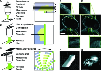
Figure 5. Fast confocal microscopy and scanning artifacts.
(A)–(C) Confocal microscopy rejects out of focus light via a pinhole aperture that is confocal with the imaging plane. (A) Point-scanning confocal microscopes require raster scanning the focused light over the whole sample to acquire an image. (B) By using light shaped to a line and a slit instead of a pinhole, superior acquisition speeds can be achieved since scanning is only required in a single direction. (C) Other ways to increase acquisition speed over point scanning is by use of multiple focused light points and pinholes, e.g., arranged on a spinning disc. (D) While point scanners provide high spatial resolution and axial selectivity (detail, top) fast moving structures such as the heart in this 30 h post fertilization (hpf) old zebrafish embryo (BODIPY FL fluorescent dye) cannot be captured with sufficient accuracy (detail, bottom). Scale bar is 100 μm. (E) With frame-rates 10–100 fold more rapid than those of point-scanning microscopes, line-scanning confocal microscopes can capture the actual structure of the beating heart (detail, bottom), while keeping good spatial resolution and optical sectioning ability (detail, top). (F) Three-dimensional cartoon of the heart-tube and (G) as measured by successively imaging planes along the Z direction of the heart in a 30 hpf transgenic Tg(cmlc2:EGFP) zebrafish. Similarly to panel D, during the time it takes for scanning along Z (2–3 s), the heart has time to beat five times resulting in a corrupted image [see also Supplementary movie (EPAPS)]. More sophisticated techniques are required to capture the actual geometry of the heart tube. Grid spacing is 20 μm.

