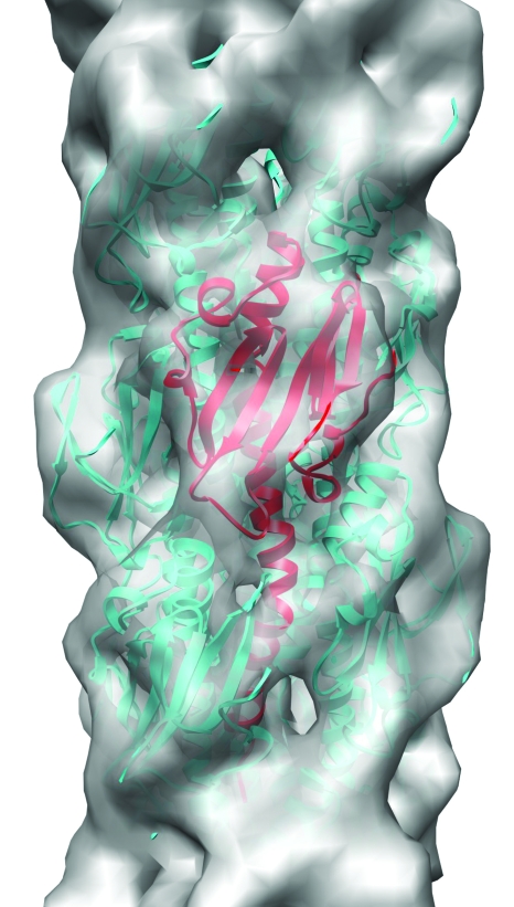
Figure 3. The type IV pilus has been reconstructed at ∼12 Å resolution from electron cryomicrographs ( Craig et al., 2006 ) and is shown as a gray surface.
This resolution provides a unique fit for the crystal structure of the component pilin (one subunit shown in red ribbons, while the surrounding subunits are shown in cyan).

