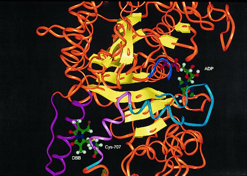
Figure 3.

Environment of bound ADP and emplaced dibromobimane. Except for select, otherwise specified, portions, the “trace” is rendered as an orange “oval,” and the β-strands as yellow arrows. The helix that connects Cys-707 and Cys-697 is in light magenta; it connects via a helix and loop (both cyan) to the third β-strand of the β-sheet. The “P-loop” (blue) of the ADP binding site connects to the fourth strand of this β-sheet. The emplaced dibromobimane, shown as attached only to Cys-707, interacts with a helix and loop (both magenta) by steric and hydrophobic contacts. This picture was created from PDB code, 1 mmd, as MMD.ADP.BeFx. ADP placement was obtained by superimposition of the trace of MMD.ADP.BeFx on the trace of MMD.DBB. Sequence numbers are those of rabbit myosin.
