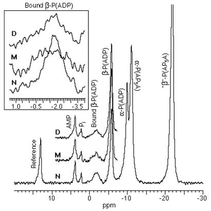
Figure 7.
The 31P chemical shift spectra of MgADP interacting with myosin S1 (in various states of derivatization) in the presence of a reference substance and an adenylate kinase inhibitor at 0°C, pH 7; N, native; M, derivatized with monobromobimane; D, derivatized with dibromobimane. (Inset) The −2-ppm peaks have been isolated and enlarged to show changes resulting from derivatization. Note that N shows no doublet structure but clearly generates a peak (see refs. 11–13).

