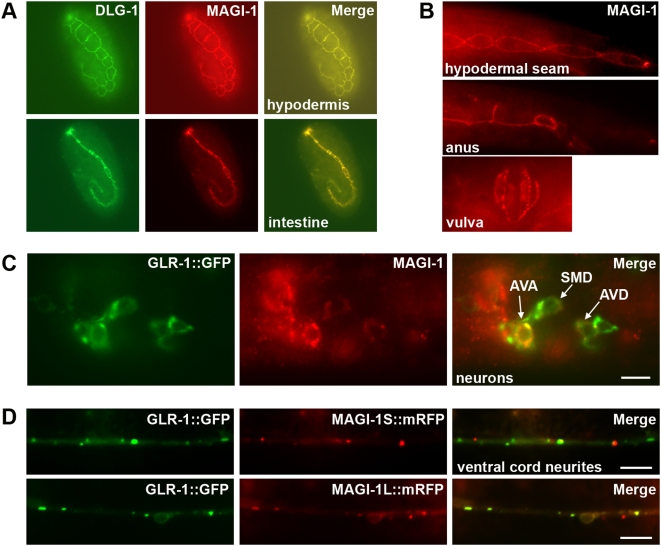
Figure 2. MAGI-1 is localized to adherens junctions and synapses.
(A) As detected by fluorescence immunohistochemistry, MAGI-1 (red) co-localizes with DLG-1 (green) in a sub-set of hypodermal tissue (top) and intestine (bottom) during development. Shown in tadpole stage embryos. (B) MAGI-1 persists in a subset of epidermal tissues, including the hypodermal seam cells, the intestine (not shown), and the anus throughout development. An early larval stage is shown, anterior to the left, ventral side down. MAGI-1 is also localized to junctions in the developing and fully formed vulva (ventral aspect). (C) MAGI-1 and GLR-1::GFP were detected by fluorescence immunohistochemistry. Lateral view of the head showing MAGI-1 (red) labeling GLR-1::GFP-expressing cells (green). Anterior is left and dorsal is up. MAGI-1 was detected in the neurons AVA, AVD, and SMD (indicated by white arrows), as well as RMD and RMDV (not shown). (D) GFP and mRFP fluorescence detected in live animals. Ventral view of interneurons expressing GLR-1::GFP (green) and a C-terminal mRFP fusion (red) to either the short (MAGI-1S, top panels) or long (MAGI-1L, bottom panels) isoform of MAGI-1 (expressed from the glr-1 promoter). Anterior is to the left. Bars, 5 µm.

