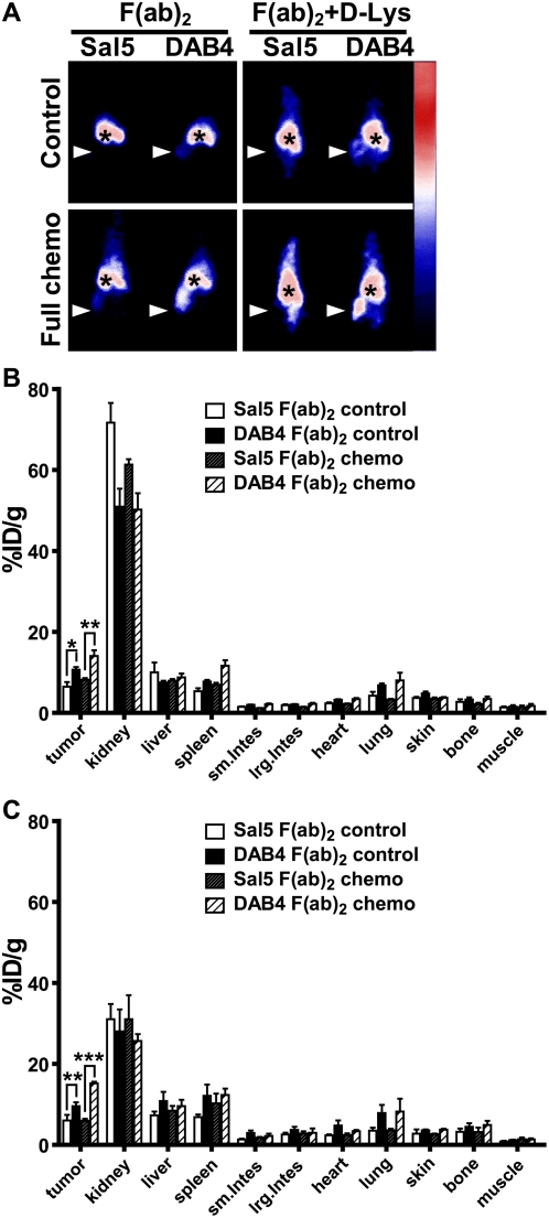
Figure 6. Imaging and biodistribution of antibody fragments.
DOTA-conjugated F(ab)2 fragments of Sal5 or DAB4 were 111In-labeled and given i.v.i. to untreated (control) or treated EL4 tumor-bearing mice 24 h after full-dose chemotherapy. Some mice receiving F(ab)2 fragments also received regular D-lysine injections. Mice were imaged with a gamma camera 24 h after radioligand administration, and then killed to measure gamma counts in tumors and normal organs. A, shown are representative gamma camera images of n = 3 mice. Accumulation of radioligand is shown as mean %ID/g±SEM (n = 3/group) for F(ab)2 forms of Sal5 and DAB4 B, without or C, with co-injection of D-lysine at 24 h (* P<0.05, ** P<0.01, *** P<0.001 for DAB4 versus Sal5). Antigen-specific and chemotherapy-dependent tumor accumulation of DAB4 occurred independently of the Fc fragment.

