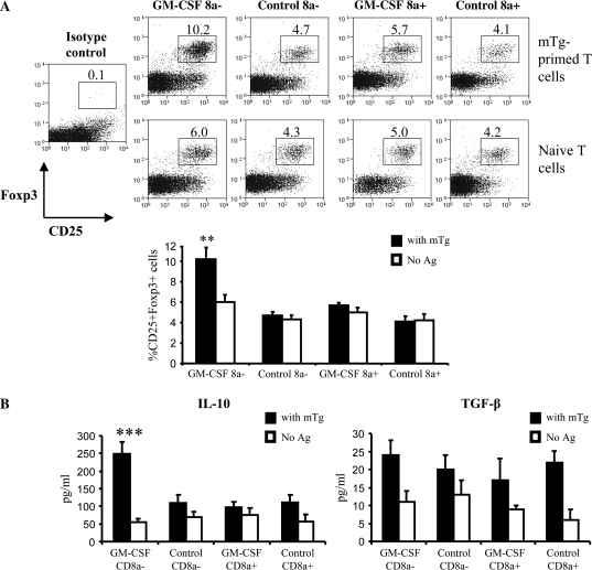
Fig. 3.
DCs from GM-CSF-treated mice cause expansion of Foxp3+ IL-10-secreting T cells in vitro: CBA/J mice (5 mice per group) were treated with GM-CSF or PBS for five consecutive days starting on days 1 and 15. CD11c+ DCs isolated on day 21 from pooled splenocytes of GM-CSF-treated or control mice were further sorted into CD8a+ and CD8a− populations as described above. CD8a+ or CD8a− DCs (2 × 10⁴ cells) were co-cultured with mTg-primed or naive CD4+ splenic T cells (purified by using CD4-labeled magnetic beads) at a 1:1 ratio in 96-well round-bottom plates. DC–T cell cultures were pulsed with 50 μg ml−1 of mTg. (A) After 6 days in culture, CD4+T cells were analyzed for Foxp3 expression along with CD25 by flow cytometry. Representative scatterplots (upper panels) and mean ± SD values of CD25+Foxp3+ cells from three independent experiments carried in triplicate (lower panel) are shown. (B) IL-10 and TGF-β levels in supernatants from 48-h culture were quantified by ELISA. Results shown are mean ± SD values from three independent experiments carried out in triplicates. One-way analysis of variance was used to determine statistical significance. ** P < 0.01.

