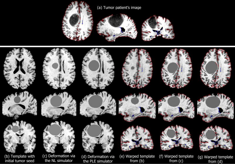
Fig. 2.
Registration example of a normal template to a brain tumor image. The brain tumor image, which corresponds to patient 3 from Tables I and II is shown in (a). The template with the initial tumor seed is shown in (b). The template with simulated tumor using (c) the NL simulator and (d) the PLE simulator is registered as shown in (f) and (g) respectively. The registration of (b) to (a) without the application of any biomechanical model of mass effect is shown in (e). The colored curves in (e–g) represent the edges of the patient’s image, overlaid on the warped template.

