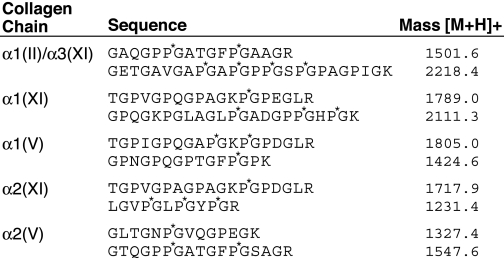
TABLE 1.
Representative tryptic peptides identified by in-gel trypsin digestion and mass spectrometry
The individual collagen bands on SDS-PAGE stained by Coomassie Brilliant Blue shown in Fig. 6 were cut out and digested in-gel by trypsin and analyzed by mass spectrometry. P*, 4-hydroxyproline residue; K*, hydroxylysine residue.

