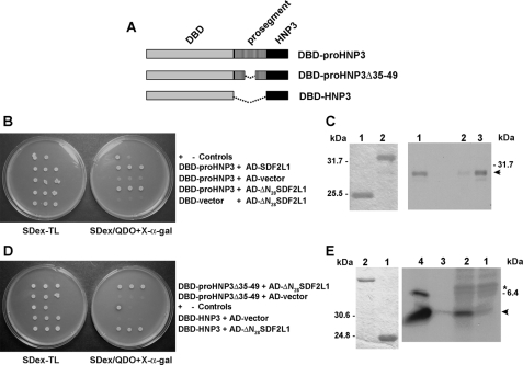
FIGURE 4.
SDF2L1 interacts with proHNP3 and the mature HNP3 sequence is sufficient for this interaction. A, schematic representations of the DBD-fusion constructs used in this figure. B, spot tests were performed using three colonies of AH109 cells transformed with plasmids DBD-proHNP3 and AD-SDF2L1, DBD-proHNP3, and AD-ΔN28SDF2L1, and the controls: DBD-proHNP3 and AD-vector, and DBD-vector and AD-ΔN28SDF2L1 as indicated in the figure. C, In vitro interaction between proHNP3 and SDF2L1. Left panel, SDS-tricine PAGE of GST (lane 1) and GST-proHNP3 (lane 2) purified from yeast extracts with glutathione-Sepharose beads and then Coomassie Blue-stained. Right panel, GST (lane 2) and GST-proHNP3 columns (lane 3) were incubated with [35S]HA-SDF2L1. Bound proteins were analyzed by autoradiography. Lane 1, input [35S]HA-SDF2L1 (arrowhead). D, spot tests of AH109 cells transformed with plasmids listed (Table 1). E, in vitro interaction between HNPs and SDF2L1; left panel, Coomassie Blue-stained SDS-Tricine PAGE of GST (lane 1) and GST-ΔN28SDF2L1 (lane 2) prepared as in C above. Right panel, GST (lane 1) and GST-ΔN28SDF2L1 (lane 2) columns incubated with metabolically labeled HL60 extracts. Bound proteins were analyzed by autoradiography. Control immunoprecipitations with labeled HL60 extracts and protein A-Sepharose beads and either rabbit preimmune IgG (lane 3) or α-HNP IgG (lane 4). The arrowhead indicates 35S-labeled HNP, and the asterisk indicates the precursor, proHNP.

