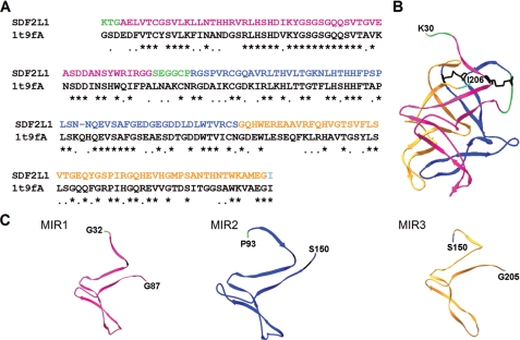
FIGURE 6.
Homology model of SDF2L1 and the structure of its MIR domains. SWISS-MODEL was used to construct a three-dimensional structure of SDF2L1 that includes residues 30–206. The MIR1, MIR2, and MIR3 domains are represented in magenta, blue, and orange, respectively. The N-terminal sequence and the sequence between MIR1 and -2 are green, and the C-terminal residue is shown in cyan. The N- and C-terminal residues are indicated in the structures. A, alignment of rhesus SDF2L1 residues 30–206 with the chain A of 1t9f.PDB sequence (1t9fA). B, predicted structure of SDF2L1 displayed using the SWISS-PDBviewer. The Cys residues and the putative disulfide linkages in this structure are shown in black. C, structures of individual MIR1, -2, and -3 domains; an extra N-terminal residue was also included to indicate the sequence orientation of each domains.

