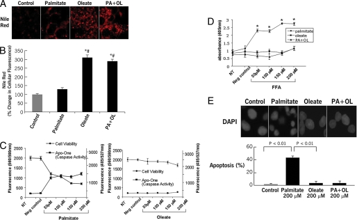
FIGURE 1.
Triglyceride accumulation and cell viability in FFA-treated hepatocytes. Isolated hepatocytes from C57BL/6 mice were incubated in the presence or absence of various concentrations of FFA with different degrees of saturation for up 24 h. A, triglyceride accumulation was determined by staining with Nile Red in fixed cells and (B) quantitated by digitized fluorescence microscopy. C, cell viability and caspase activation were assessed by CellTiter Blue and Apo-ONE Homogeneous Caspase 3/7 fluorometric assays, respectively. Finally, apoptotic cell death was quantified by an ELISA cell death assay (D) and using the DNA-binding dye 4′,6-diamidino-2-phenylindole (DAPI) coupled to fluorescence microscopy (E). Results are expressed as mean ± S.D. from three independent experiments. *, p < 0.05 compared with controls. #, p < 0.05 compared with palmitate-treated cells.

