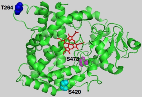
FIGURE 7.
PyMol depiction of CYP3A4 phosphorylatable Thr264, Ser420, and Ser478 residues. The CYP3A4 crystal structure (48) was used as the template, depicting the prosthetic heme in red, phosphorylatable residues Thr264 in blue, Ser420 in cyan, and Ser478 in magenta.

