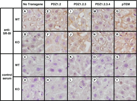
FIGURE 3.
Immunohistochemical analysis of hepatic SR-BI in WT and PDZK1 KO nontransgenic (A-D), PDZ1.2 transgenic (E-H), PDZ1.2.3 transgenic (I-L), PDZ1.2.3.4 transgenic (M-P), and pTEM transgenic (Q-T) mice. Livers from mice with the indicated genotypes and transgenes were fixed, frozen, and sectioned. The sections (5 μm) were then incubated with either polyclonal anti-SR-BI antiserum (top two rows) or control rabbit serum (bottom two rows) followed by a biotinylated anti-rabbit IgG secondary antibody and then stained using immunoperoxidase reagents and hematoxylin counterstaining. (Magnification, ×1200.) The samples from transgenic animals are from the following founder lines: 836 (E and G), 833 (F and H), 644 (I-L), 1163 (M-P), and 6656 (Q-T). Images are representative of the staining patterns observed in all founder lines for each transgene.

