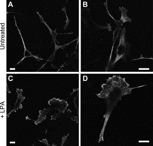
FIGURE 7.
LPA-stimulated MDA-MB-231 cells display high PKA activity at the leading edge. MDA-MB-231 cells were plated on collagen-coated coverslips and then left untreated (A and B) or treated with 100 nm LPA for 15 min (C and D). Cells were then immunostained for phospho-PKA substrates as described under “Experimental Procedures.” Representative confocal images for each condition are shown at two different magnifications. The bar in each panel indicates 10 μm.

