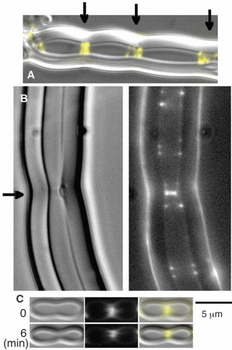
Fig. 3.
FtsZ-mts and GTP were mixed with liposomes. Although the FtsZ-mts was initially outside the liposomes, some tubular, multilamellar liposomes were formed that enclosed FtsZ-mts and GTP. The FtsZ assembled into Z rings in these tubular liposomes. (A) A liposome with three bright Z rings, each centered on a constriction. The fluorescent FtsZ is shown in yellow, superimposed on the differential interference contrast image of the liposome. Arrows indicate Z rings. (B) The bright Z ring near the middle (indicated by the arrow) is forming a visible constriction and on the right side appears to have detached some inner layers of the multilamellar wall. (C) A liposome is shown here with a visible constriction at the Z ring when first observed, 5 to 10 min after making the specimen. Six minutes later the constriction has narrowed markedly. See movie S1 for a 10-min series showing Z rings coalescing into brighter ones, which generate constrictions.

