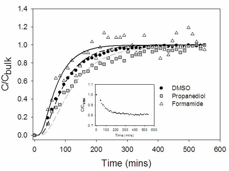
Figure 4.
Representative concentration profiles of permeating half strength VS55 (1.55 M DMSO, 1.55 M formamide, 1.05 M 1,2 propanediol) into articular cartilage. Points are experimental measurements from localized 1H NMR spectroscopy from a 1×1×1 mm3 volume of interest (VOI) positioned in the center of the plug. Lines are best fit model simulations using the effective diffusivity of each component as the only adjustable parameter. Water was also detected in the 1H NMR spectra (Figure 3). As the tissue is exposed to a hypertonic CPA solution, water leaves the tissue simultaneously as the influx of the VS55 components occurs. The inset shows the decline in water concentration in the VOI, normalized to the initial water concentration (C/Cinitial), vs. time.

