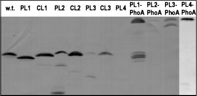
Figure 2.
Protein expression and stability. Circularly permuted variants were visualized with mAbs against the IIBGlc domain on a Western blot of membrane preparations. PL2, PL3, PL1-PhoA, PL2-PhoA, and PL3-PhoA are partially degraded. A total of 100 μg of membrane protein was loaded per lane. w.t., wild-type.

