
Figure 7.
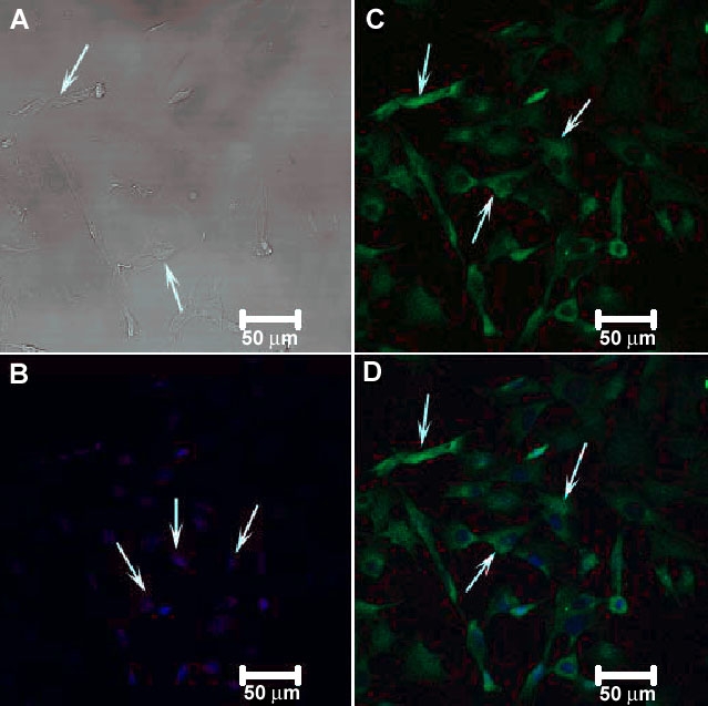
Confocal immunocytochemical localization of complement factor H (CFH) in RGC-5. The DIC image is shown in (A) and the nuclear staining by Hoechst stain is shown in (B). Arrows in A and B depict the cells and the nuclei of the RGC-5 cells, respectively. Complement factor H (CFH) in green (arrows in C). The merged images of these RGC-5 cells are shown in D (arrows). CFH was expressed in the cytoplasm (arrows in B and D).