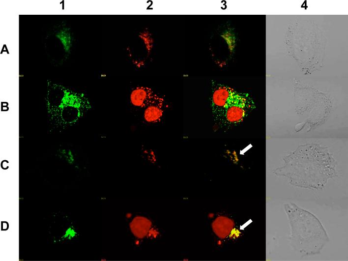
Figure 9. Co-localization of 623 with endosomal pathway markers.
RPA-623-Tamra (100 nM) prepared by conjugating a HSA surface thiol group to a cysteine instead of Alexa 488, was co-incubated with (A) Transferrin-Alexa 488 (200 nM) for 2 h, (B) Transferrin-Alexa 488 (100 nM) for 24 h, (C) Dextran-Alexa-488 (2 uM) for 2 h, (D) Dextran-Alexa-488 (2 uM) for 24 h. Live cells were observed for 1) Alexa-488, 2) Tamra, 3) Merged images of Alexa-488 and Tamra and 4) differential interference contrast (DIC) image using an Olympus confocal fluorescence microscope as described in experimental procedures. Arrows represent colocalization of Alexa 488 and Tamra fluorophores.

