
Figure 2.
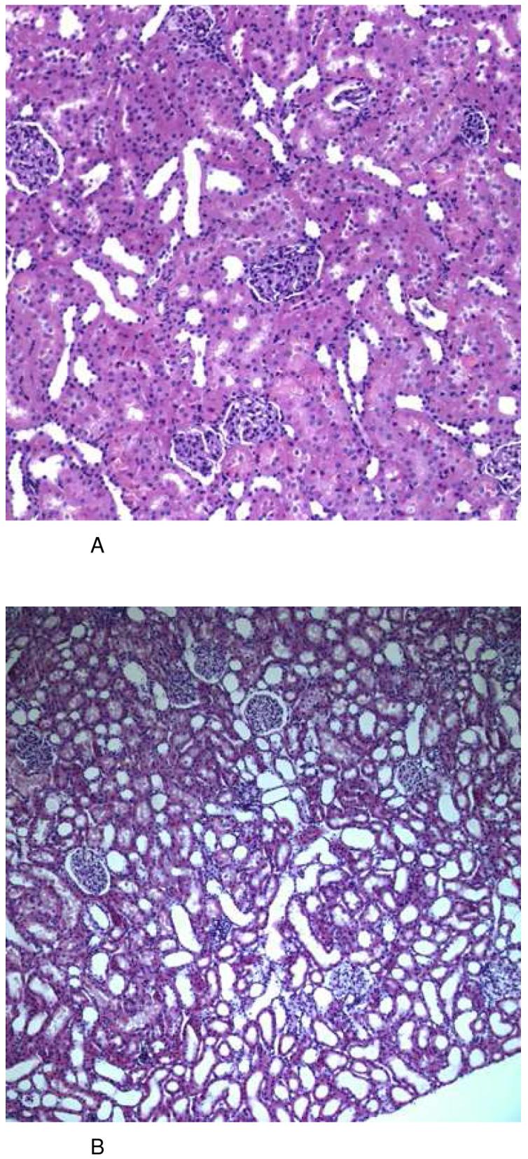
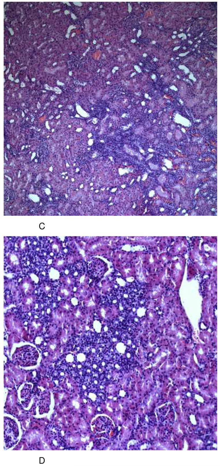
Representative histology (HE stain) of kidney tissues from the different treatment groups at day 28 (total number of tissue samples evaluated: n=6/ treatment group, all histologies (A-C): magnification 10×22, (D): magnification 20×22).
After 6 days of treatment, none of the groups revealed any significant histological changes in comparison to kidneys from vehicle-treated controls (A). After treatment with sirolimus (1 mg/kg/day), kidneys revealed alterations of the tubular system appearing as severe atrophy and dilation. These changes were reproducible and clearly detectable in every animal (B). Following long-term treatment with cyclosporine (10 mg/kg/day), kidneys showed a patchy alteration caused again by mild tubular atrophy (shrinking of the proximal tubular cytoplasm) plus atrophy and luminal dilation of the distal tubulus system. As another typical finding, there was a most significant presence of micro- and macrovesicular occlusion bodies in almost all tubular epithelial cells (C). These changes were similar and much more pronounced in the group receiving a combination treatment protocol of sirolimus and cyclosporine (D).
Groups: con: vehicle-treated controls, CsA10: 10 mg/kg/day cyclosporine, CsA25: 25 mg/kg/day cyclosporine, Rapa1: 1 mg/kg/day sirolimus, CsA10/Rapa1: co-administration of 10 mg/ kg/ day cyclosporine and 1 mg/kg/day sirolimus, CsA25/Rapa1: co-administration of 25 mg/kg/day cyclosporine and 1 mg/kg/day sirolimus.