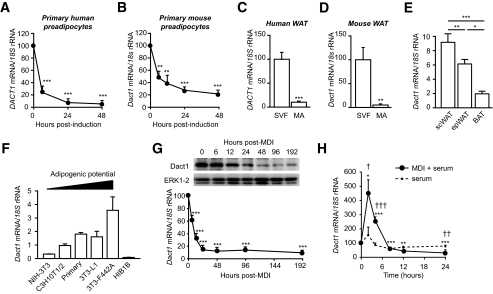
FIG. 1.
DACT1 gene expression is downregulated in early adipogenesis and is found primarily the SVF of human and mouse adipose tissue. Human DACT1 (A) and mouse Dact1 (B) mRNA levels were measured using real-time RT-PCR at the indicated time points after induction of differentiation. **P < 0.01, ***P < 0.001 vs. time 0, n = 7 experiments performed in duplicate. DACT1 and Dact1 mRNA levels were measured in stroma-vascular cells (SVF) and mature adipocytes (MA) from human subcutaneous WAT (C) and mouse epididymal WAT (D). ***P < 0.001 vs. SVF, n = 3 replicate experiments performed in duplicate. n = 3 experiments performed in duplicate. E: Normalized Dact1 mRNA levels were measured in epididydimal (epWAT), subcutaneous WAT (scWAT), and interscapular BAT of 10-week-old C57BL/6J. *P < 0.05, **P < 0.01, ***P < 0.001, n = 3 experiments performed in duplicate. F: Dact1 mRNA levels, were measured in different cell types: fibroblast (NIH3T3), mesenchymal stem cells (C3H10T1/2), white primary preadipocytes, white preadipocyte cell lines (3T3–L1 and 3T3–F442A), and brown preadipocyte cell line (HIB1B), n = 3–4 experiments performed in duplicate. G: Dact1 mRNA levels were measured at the indicated hours after induction (with MDI) of 3T3–L1 preadipocyte differentiation. ***P < 0.001 vs. time 0, n = 4 experiments performed in duplicate. Whole-cell protein lysates were extracted at the indicated days of differentiation from 3T3 L1 cells and analyzed by immunoblotting. Representative immunoblots of Dact1 and extracellular signal–related kinase (ERK)1/2 (loading control) from three separate experiments are shown. H: Dact1 mRNA levels were measured at the indicated hours after induction of 3T3–L1 preadipocytes with either control medium (serum) or MDI. *P < 0.05, **P < 0.01, and ***P < 0.001 vs. time 0 for MDI; †P < 0.05, ††P < 0.01, and †††P < 0.001 vs. serum, n = 3–4 experiments performed in duplicate.

