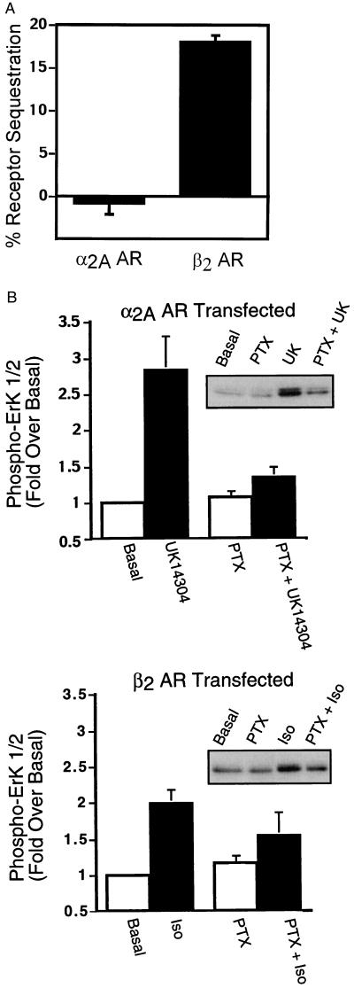
Figure 1.
Agonist-promoted α2A AR and β2 AR sequestration and ERK 1/2 phosphorylation in COS-7 cells. (A) COS-7 cells transiently expressing either HA-epitope tagged α2A ARs or Flag epitope-tagged β2 ARs were serum-starved overnight and exposed to UK14304 (10 μM) or isoproterenol (10 μM), respectively, for 30 min at 37°C. Cell-surface receptors were labeled with an 12CA5 monoclonal antibody or an M2 Flag monoclonal antibody, by using FITC-conjugated goat anti-mouse IgG as the secondary antibody. Receptor sequestration, quantified as the percent loss of cell-surface fluorescence in agonist-treated cells, was measured by using flow cytometry. The data are expressed as the mean ± SEM of four independent experiments performed in triplicate. (B) Appropriately transfected COS-7 cells were serum-starved overnight in the presence or absence of pertussis toxin (100 ng/ml) before stimulation with 1 μM UK14304 (Upper) or 1 μM isoproterenol (Lower) for 5 min. Aliquots of whole-cell lysate (approximately 30 μg of protein per lane) were resolved by SDS/PAGE, and ERK 1/2 phosphorylation was detected by protein immunoblotting by using rabbit polyclonal phospho-MAPK-specific IgG. Data are expressed as the fold ERK 1/2 phosphorylation over the basal value in appropriately transfected cells. The data are expressed as the mean ± SEM of three independent experiments.

