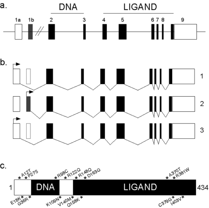
Figure 1. Structure of SXR gene, protein and mRNA isoforms.
a) The genomic structure of SXR and its three splice variants. The protein coding regions are depicted as filled boxes and the untranslated 5’ and 3’ regions are shown as white boxes. The horizontal line represents introns. b) The structure of splice variants of SXR. Variant 1 originates from exon 1a and gives rise to proteins 1l and 1s through the use of the alternative initiation codons shown by arrows. Variant 2 originates from exon 1b and it makes the longest protein. Variant 3 represents an in-frame deletion of 111bp at the 5’ end of exon 5 (shown by white box). The arrows depict translation initiation codon. c) Single nucleotide polymorphisms (SNPs) in SXR. Fifteen known non-synonymous SNPs of SXR are represented by * and arranged according to their position on the wild-type (variant 1l) SXR protein. The DNA and ligand binding domains of the protein are shown in black boxes.

