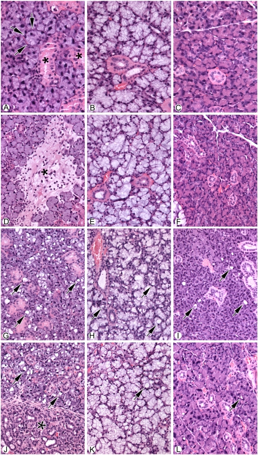
Figure 5. Histological analysis of salivary glands from normal and irradiated salivary glands from untreated and IGF1-treated mice.
A) Normal submandibular salivary gland showing basic structure of acini (arrows) and ducts (asterisk). B) Normal sublingual salivary gland. C) Normal parotid gland. D) Submandibular salivary gland thirty days following exposure to 5 Gy radiation. Note area of focal fibrosis and associated inflammatory cells (asterisk). E) Sublingual salivary gland after thirty days following exposure to 5 Gy radiation. No significant morphological change is seen. F) Parotid salivary gland after exposure to radiation. No Significant morphological change is seen. G) Submandibular gland thirty days following injection with 5 ug IGF1. Note the prominent vacuolization of the glandular acini (Arrows). H) Sublingual gland thirty days following injection with 5 ug IGF1 introduction. Note the prominent vacuolization of the glandular acini (Arrows). I) Parotid gland thirty days following injection with 5 ug IGF1. Note the mildly increased vacuolization of the glandular acini, although less than that observed in either the submandibular or sublingual salivary glands (Arrows). J) Submandibular gland thirty days following injection with 5 ug IGF1 immediately prior exposure to 5 Gy radiation. The left part of the photograph (*) shows atrophy of the acini with mild chronic inflammation. The right side of the picture shows increased vacuolization in the viable acini (arrows). K) Sublingual gland thirty days following injection with 5 ug IGF1 immediately prior to exposure to 5 Gy radiation. Increased vacuolization is noted, however, no significant atrophy is seen. L) Parotid gland thirty days following injection with 5 ug IGF1 immediately prior to exposure to 5 Gy radiation. Note the mildly increased vacuolization with no significant atrophy. For Panels A–C, the magnification is 40×, while the magnification D–L is 20×.

