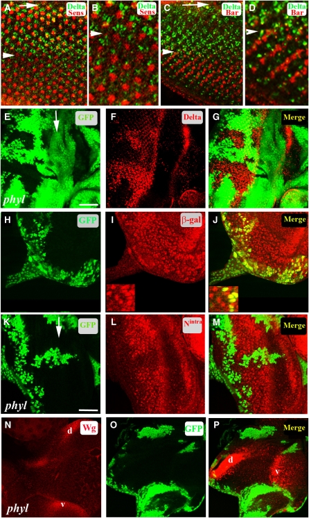
Figure 1.
Phyl-mediated downregulation of Dl, Notch and Wg in the developing Drosophila eye disc. Arrows mark the morphogenetic furrow (MF). Arrowheads mark eight columns posterior to the furrow. (A–D) Temporal regulation of Dl expression in R cells. (A) Dl expression in the third instar eye disc. Dl (green) in R cells, Senseless (red) marking the R8 cell in each cluster. Dl expression is downregulated at about column 8 (arrowhead) posterior (down) to the MF. (B) A high magnification view of (A) near column 8 (arrowhead) showing downregulation of Dl (green) posterior (down) to column 8. (C) The expression of BarH1 (red) in R1 and R6 initiates at around column 8 (arrowhead) at the time when the expression of Dl (green) is downregulated in R cells. (D) A high magnification view of (C) near column 8 (arrowhead) showing downregulation of Dl posterior to column 8. (E–G) In a disc containing a phyl clone (non-green, E), Dl is overexpressed in the mutant tissue (F, red). The merged panel is shown in (G). (H–J) In a disc containing a phyl clone (non-green, H) in the background of Dl-lacZ, the level of β-galactosidase expression (I, red nuclei) remains unaltered in mutant cells. A merged panel (J) shows no difference in Dl-lacZ expression in cells mutant for phyl when compared with adjacent wild-type cells; shown in higher-magnification insets (bottom left corner of I and J). (K–M) In a disc containing a phyl clone (non-green, K), increased amount of Notch detected by an antibody against the intracellular domain of Notch (red, L) is seen in the phyl mutant cells when compared with adjacent wild-type cells. A merged image is shown in (M). (N–P) In a wild-type third instar eye disc, Wg is expressed on the dorsal (d) and ventral (v) edges of the eye disc (N). In a disc containing large phyl mutant clones (non-green in O), increased amount of Wg is seen in the mutant tissue along the dorsal/ventral edges (red in P).

