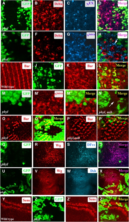
Figure 2.
Processed forms of Notch and activated version of Wg signalling complex accumulate in phyl mutant cells. (A–H) phyl mutant clones in pupal eye discs. (A–D) In phyl mutant cells (non-green in A and D), Dl (red, B) co-localizes with extracellular domain of Notch, Nex (blue, C). The merged panel is shown in (D). (E–H) In a pupal eye disc containing phyl mutant clones (non-green in E and H) increased level of Dl is seen in mutant tissue (red, F). The same disc co-stained with an antibody directed against the intracellular domain of Notch shows an accumulation of Nintra in adjacent pigment cells (blue, G). The merged panel shows this lack of co-localization (arrow, H). (I–L) In wild-type mid-pupal eye disc, Bar (red, I) is expressed in primary pigment cells. Mid-pupal eye disc containing phyl mutant clones (non green in J) shows an increase in the number of Bar-expressing cells (red, K). The merged panel is shown in (L). (M, N) Increased Nintra level in phyl mutant cells requires Dl signalling clones of phyl mutant tissue in third instar eye disc (non green in M) stained for Nintra (red, M′). The merged panel (M″) shows that only non-green mutant cells accumulate Notch. In phyl mib1 double-mutant clones, Nintra does not accumulate in the mutant tissue (arrow, compare M′ with N). (O–P′) Loss of Notch signalling rescues phyl mutant phenotype. phyl mutant clones (non-green, O′) show a loss of Bar-expressing R1 and R6 cell types (red, O). In phyl, mib1 double-mutant clones (non-green in P′), the phyl mutant phenotype is rescued and Bar-expressing R1 and R6 cell types are seen in the mutant tissue (red, P). (Q–T) Third instar eye disc containing phyl mutant (non-green in Q) clones shows increased levels of Wg protein (red, R). The same disc was also stained with DFrz (blue, S), showing co-localization of Wg with Dfrz (pink, T). (U–X) Third instar eye disc containing phyl mutant clones (non-green in U) shows increased levels of Wg protein (red, V). The same disc was also stained with Dsh (blue, W), showing co-localization of Wg with Dsh (pink, X). (Y–Z″) In wild-type third instar wing disc, Senseless is expressed in cells straddling the D/V boundary (red, Y). phyl mutant clones in the wing disc generated using ubx-flp (non green in Z) show an expansion of Senseless expression domain (red, Z′). The merged panel is shown in (Z″).

