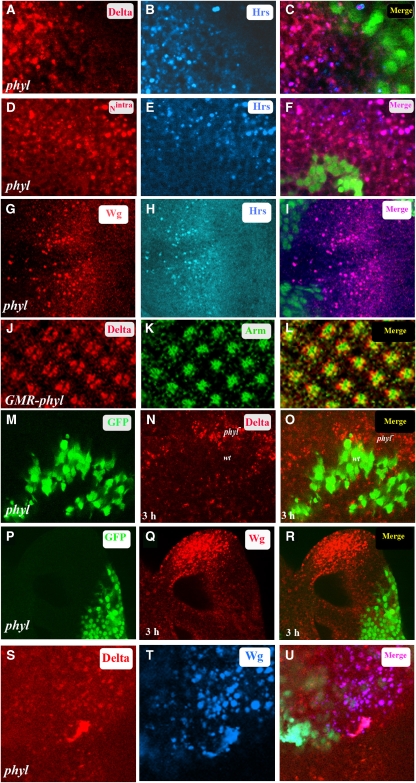
Figure 3.
Defects in endocytic trafficking of Dl, Nintra and Wg in phyl mutant cells. (A–I) phyl mutant clones in third instar eye disc. GFP marks wild-type cells and non-GFP cells are mutant for phyl. (A–C) In phyl mutant cells (non-green in C), Dl (red, A) co-localizes with HRS, which marks endocytic vesicles (blue, B). The merged panel is shown in (C). (D–F) In phyl mutant cells (non-green in F), Nintra (red, D) co-localizes with HRS (blue, E). The merged panel (F) shows this co-localization (pink, F). (G–I) In phyl mutant cells (non-green in I), Wg (red, G) co-localizes with HRS (blue, H). The merged panel shows this co-localization (pink, I). (J–L) GMR-phyl third instar eye disc stained for Dl (red, J) and Arm marking the apical tips of the R cells (green, K). The merged panel (L) shows co-localization of Dl and Arm in the plasma membrane (yellow, L). (M–R) Block in endocytic trafficking of Dl and Wg in phyl mutant cells. Live labelling followed by a 3 h chase in third instar eye disc containing a phyl mutant clone (non-green in M and P) stained for Dl (red in N) or Wg (red in Q). Following the chase, Dl and Wg proteins were detected only in phyl mutant cells and not in adjacent wild-type cells where they have been degraded (O and R). (S–U) Dl and Wg co-localize to the same endocytic vesicles. Third instar eye disc containing clones of phyl (non-green tissue in U) stained with Dl (red, S) and Wg (blue, T). The merged panel (U) shows co-localization of Dl and Wg in the endocytic vesicles.

