Figure 1.
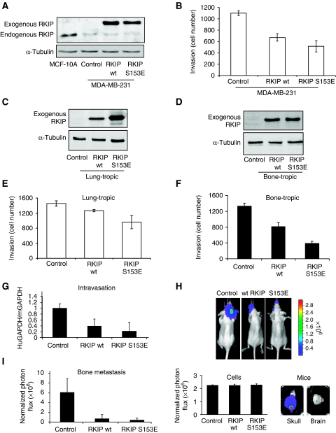
RKIP regulates breast cancer invasion and metastasis. (A) RKIP is expressed in MCF10A mammary gland and depleted in metastatic MDA-MB-231 cells. MDA-MB-231 cells were stably transduced with wt or S153E RKIP and the lysates immunoblotted with anti-RKIP or anti-tubulin antibody. (B) Wt and S153E RKIP inhibit invasion of MDA-MB-231 cells. Cells were assayed for invasion as described in Materials and methods. Results represent the mean±s.e. for four independent samples (P<0.001 for wt and P=0.002 for S153E RKIP relative to Control). (C, D) Expression of wt and S153E RKIP in lung-tropic (4175) or bone-tropic (1833) breast cancer cells. Cells were stably transduced with wt or S153E RKIP and the lysates were immunoblotted with anti-RKIP or anti-tubulin antibody. All lanes in (C) come from the same gel, but one lane after sample 2 was omitted leading to a composite figure. (E, F) Wt and S153E RKIP inhibit invasion of lung-tropic (4175) or bone-tropic (1833) cells. The 1833 cells stably expressing control vector, wt RKIP, or S153E RKIP were assayed for invasion as described in Materials and methods. Results represent the mean±s.e. for four independent samples (4175) or mean±s.d. for three samples (1833) (P<0.05 for wt RKIP and P<0.05 for S153E RKIP relative to control 4175 cells; P=0.02 for wt RKIP and P<0.001 for S153E RKIP relative to control 1833 cells). (G) Wt and S153E RKIP inhibit intravasation of bone-tropic tumour cells (1833). The 1833 cells stably expressing control vector (six mice), wt RKIP (five mice), or S153E RKIP (five mice) were injected into the mammary fat pad of mice. After 3 weeks, cells isolated from the blood were analysed for GAPDH transcripts derived from human (tumour) or mouse (control). Results represent the mean±s.d. for the animals (P<0.002 for wt RKIP and P<0.001 for S153E RKIP relative to control). (H, I) Wt and S153E RKIP inhibit bone metastases. The 1833 cells expressing luciferase and control vector (six mice), wt RKIP (seven mice), or S153E RKIP (seven mice) were injected into the left ventricle of the mice, and the mice were imaged for luciferase activity after 3 weeks. Representative images show that RKIP wt and S153E greatly reduced bone metastases in skull (H, upper right panel; lower right panel). The 1833 cells stably expressing luciferase have an identical luciferase reporter activity before injection into mice (H, lower left panel). Comparable regions of the mouse skulls were optically imaged and quantified. Results (I) represent the mean±s.d. for the animals (P<0.01 for wt RKIP and P<0.01 for S153E RKIP relative to Control).

