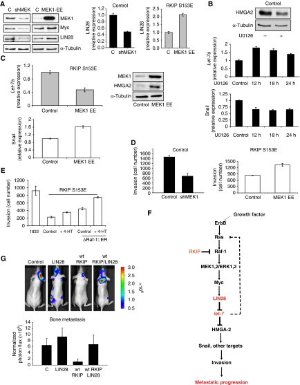
Figure 5.
RKIP regulates let-7 in part through the MAPK pathway, and LIN28 reverses the inhibition of bone metastasis by RKIP. (A) MEK1 regulates LIN28 and Myc in 1833 cells. Constitutively, active MEK induces LIN28 and Myc. The 1833 cells expressing S153E RKIP were transfected with control vector (C) or MEK1-EE for 48 h. LIN28 was also assayed by qRT–PCR. MEK1, Myc, lIN28, and α-tubulin expression were measured by western blot. Conversely, stable depletion of MEK by lentiviral shRNA downregulates LIN28 and Myc in 1833 cells. Results represent the mean±s.d. for three samples (P=0.005 for shMEK and P=0.001 for MEK1-EE relative to Control). Immunoblot results are representative of at least three independent experiments. (B) Inhibition of MEK by U0126 induces let-7a and inhibits HMGA2 and Snail. Blot: 1833 cells treated with or without the MEK inhibitor U0126 (10 μM, 12 h) were lysed and immuoblotted with antibodies to HMGA2 or tubulin. Graphs: 1833 cells were treated with 10 μM U0126 for the indicated times, and let-7a was assayed by qRT–PCR. Snail expression was quantitated by qRT–PCR. Results represent the mean±s.d. for three samples (Let-7: P=0.001, 0.01, and 0.03 for 12, 18, and 24 h, respectively, relative to Control; Snail: P=0.01, 0.004, and 0.01 for 12, 18, and 24 h, respectively, relative to Control). Results are representative of at least three independent experiments. (C) Constitutively active MEK inhibits let-7a expression and induces HMGA2 and Snail. The 1833 cells expressing S153E RKIP were transfected with control vector or MEK1-EE for 48 h, and let-7a and Snail were assayed by qRT–PCR. Results represent the mean±s.d. for three independent samples (Let-7a: P<0.02 for MEK1-EE relative to Control; Snail: P=0.01 for MEK1-EE relative to Control). MEK1 and HMGA2 expression were measured by western blot. Results are representative of at least three independent experiments. (D) MEK regulates invasion. Left: 1833 cells expressing either vector control or shRNA for MEK1 were assayed for invasion as described in Materials and methods. Results represent the mean±s.d. for three independent samples (P=0.004 for shMEK1 relative to Control). Right: 1833 cells expressing either vector control or an expression vector for MEK1-EE were assayed for invasion as described in Materials and methods. Results represent the mean±s.d. for three independent samples (P<0.005 for MEK1-EE relative to Control). (E) Inducible Raf kinase rescues the inhibition of invasion caused by S153E RKIP. The 1833 cells expressing S153E RKIP and transfected with control vector or ΔRaf-1:ER were untreated or treated with tamoxifen (4-HT). Results represent the mean±s.e. for three independent samples (P<0.001 for S153E RKIP+ΔRaf-1:ER+HT relative to S153E RKIP Control; P<0.001 for S153E RKIP+ΔRaf-1:ER+HT relative S153E RKIP+ΔRaf-1:ER). (F) Scheme showing the mechanism for RKIP regulation of invasion and metastasis through the MAPK/LIN28/let-7 pathway. (G) LIN28 overcomes the inhibitory effect of wt RKIP on bone metastasis. The 1833 cells expressing luciferase and either control vector or wt RKIP were stably transfected with control vector or LIN28 and injected into the left ventricle of the mice (Control, six mice; LIN28, 5 mice; wt RKIP, six mice; wt RKIP+LIN28, five mice), and mice were imaged for luciferase activity after 3 weeks. Results represent the mean±s.d. for the animals (P=0.001 for wt RKIP relative to Control, and P=0.01 for wt RKIP+LIN28 relative to wt RKIP).

