Figure 3.
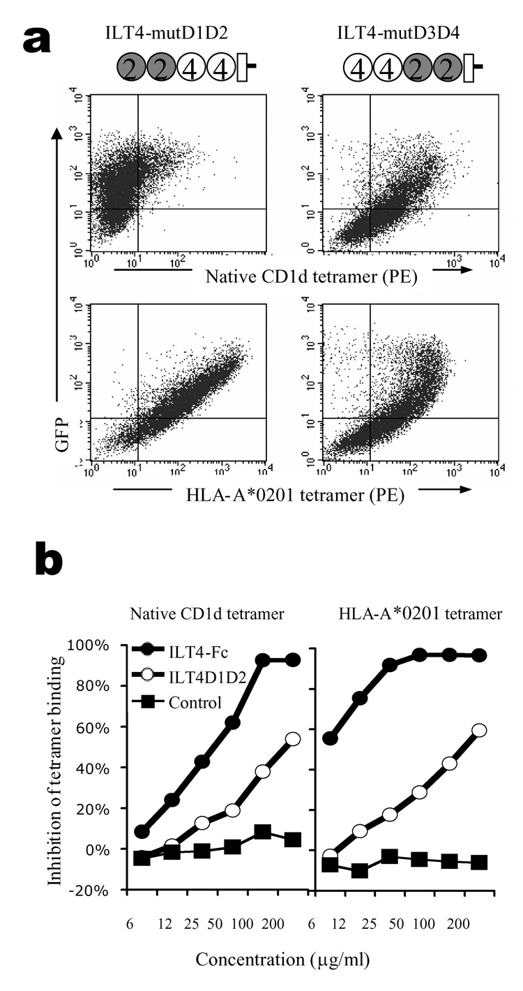
ILT4 D1–D2 are the main binding site for CD1d. (a) Binding of ILT4 mutants by CD1d. HEK293T cells transfected with GFP fusion constructs encoding ILT4 mutants were stained with PE-conjugated CD1d or HLA-A*0201 tetramers. Cartoons illustrate the structure of the constructs. Open circles represent ILT4 domains, and filled circles represent ILT2 domains. (b) Blocking of tetramer binding to ILT4-transfectants. CD1d (left panel) or HLA-A*0201 (right panel) tetramers were used to stain HEK293T-ILT4 cells in the presence of different concentrations of blocking proteins. Results from one out of three repeated experiments are shown.

