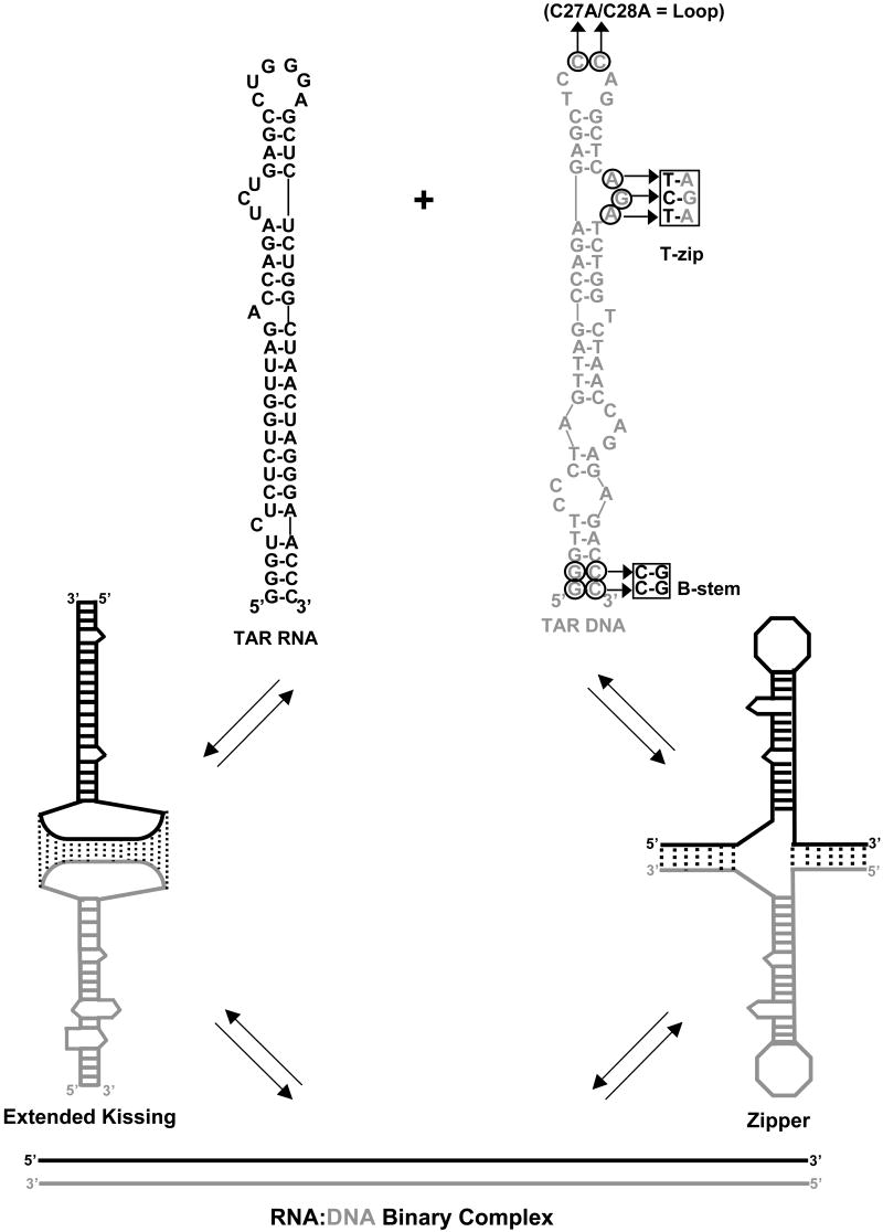
Figure 1.
Two pathways of full-length TAR RNA/DNA annealing. Top: predicted secondary structures of full-length TAR RNA (black) and TAR DNA (gray) hairpins. Sequences are derived from the HIV-1 NL4-3 isolate. The TAR RNA and DNA secondary structures were predicted by m-fold analysis51. Residues mutated in different DNA constructs are circled. Bottom: (left) loop-loop “kissing” pathway, which involves initial formation of an extended “kissing” complex followed by subsequent strand exchange to form the fully annealed duplex; (right) “zipper” pathway involving nucleation through the 3′/5′ termini resulting in formation of a zipper intermediate followed by conversion to the fully annealed duplex.

