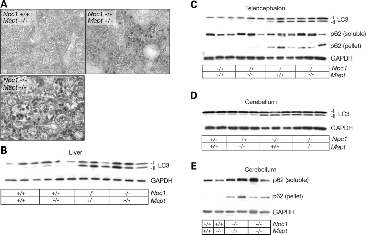
Figure 4.
LC3 and p62 expressions in NPC1 and NPC1/tau null mice. (A) Immunohistochemical staining demonstrates frequent LC3-positive vacuoles in liver of 10-week-old double null mutant (Npc1−/−; Mapt−/−) when compared with single null mutant (Npc1−/−; Mapt +/+) or wild-type mice (original magnification, 1000×). (B) Liver lysates from 10-week-old mice were probed by western blot for expression of LC3. Both double null (Npc1−/−; Mapt−/−) and NPC1 single null mice (Npc1−/−; Mapt +/+) demonstrate increased levels of LC3-II compared WITH controls. GAPDH serves as a loading control. (C–E) Lysates from the telencephalon (C) and cerebellum (D and E) of NPC1 single null, and NPC1/tau double null mice demonstrate increased levels of LC3-II and SDS-insoluble p62 compared with controls.

