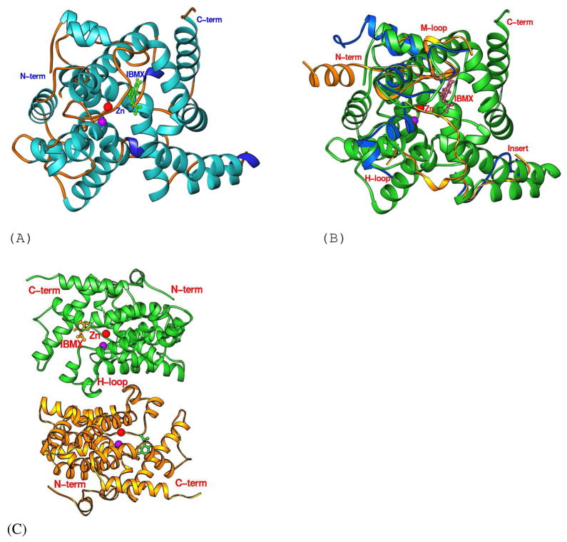
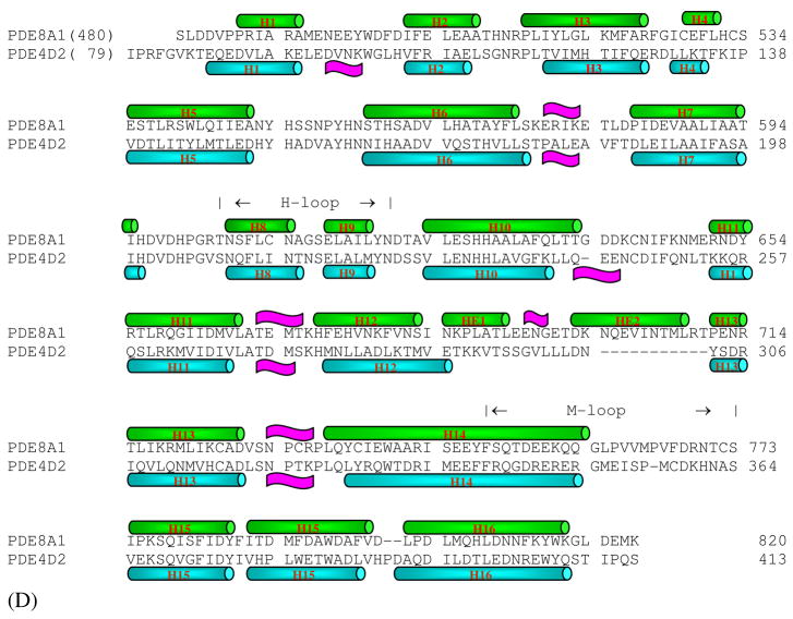
Fig. 1.
The PDE8A1 structure. (A) Ribbon diagram of the PDE8A1 catalytic domain. The purple ball represents the second divalent metal that needs to be identified. (B) Superposition of PDE8A1 (green ribbons) over PDE4D2 (blue ribbons) and PDE5A1-sildenafil (golden ribbons). The comparable portions of the PDE4D2 and PDE5A1 structures were omitted. The H-loop of PDE8A1 is similar to that of PDE4D2, but different from that of PDE5A1. (C) Dimer of PDE8A1 catalytic domain. The H-loops (residues 604-620) form the dimer interface. (D) Sequence alignment between PDE4D2 and PDE8A1.


