
Fig. 2.
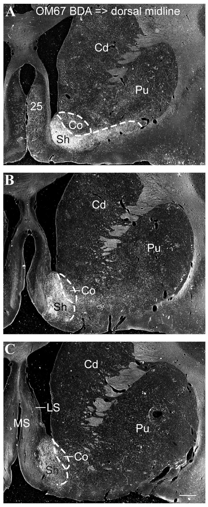
Case OM67. Darkfield photomicrographs of the striatum following a BDA injection into the dorsal midline thalamus (Fig. 1A), at three rostral-caudal levels (A-C). Labeled axons and axonal varicosities were densely concentrated in ventromedial striatum, in the shell of the accumbens nucleus. Dashed lines show approximate delineation of core and shell based on our observations and previous reports (Meredith et al., 1996; Brauer et al., 2000). Labeling can also be seen in cortical area 25 (A) and lateral septum (C) (see also Fig. 1C, D). Scale bar = 1 mm.