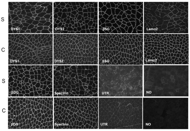
Figure 3. Immunofluorescence staining of frozen muscle biopsy sections from a dystrophic Sphynx cat showed decreased staining for laminin α-2 and increased staining for utrophin compared to a control cat.

Staining intensity with monoclonal antibodies against the rod (DYS1) and carboxy terminus (DYS2) of dystrophin, β-sarcoglycan (β-SG), β-dystroglycan (β-DG), and spectrin in the Sphynx cat (S) was similar to control (C) muscle. Staining for laminin α-2 (Lama2) was decreased while staining for utrophin (UTR) was increased in the dystrophic Sphynx cat compared to control muscle. Bar = 50 μm.
